


Dental Portable X-Ray Systems: Intraoral and Extraoral Solutions for Dental Practices
March 02, 2026 · ARRAD
Dental radiography represents fundamental diagnostic capacity in contemporary dental practice, essential for caries detection, periodontal disease assessment, dental implant planning, endodontic treatment guidance, and comprehensive oral health evaluation. Modern dental practices increasingly rely on digital radiography systems enabling rapid image acquisition, enhanced diagnostic visualization, and seamless integration with practice management software supporting treatment planning, documentation, and patient communication. Portable dental X-ray systems specifically engineered for dental applications provide flexible imaging solutions enabling comprehensive radiographic assessment directly within dental treatment environments while maintaining radiation safety standards and diagnostic quality appropriate for dental clinical decision-making.
Dental X-ray equipment differs substantially from general medical radiography systems due to dental-specific anatomic requirements, dose considerations, and clinical applications. Dental portable systems employ specialized generators, detectors, and positioning apparatus optimized for intraoral and extraoral imaging of dental structures, maxillary/mandibular alveolar bone, and associated anatomic regions relevant to dental diagnosis and treatment. These specialized systems represent distinct category within portable radiography market, serving dental practices ranging from solo practitioners to large group practices, dental schools, and pediatric dental specialists.
Intraoral Radiography: Fundamental Diagnostic Capability in Dental Practice
Intraoral radiography involves placement of small sensors or film inside patient mouths, enabling high-resolution imaging of individual teeth, tooth roots, and surrounding alveolar bone. Periapical radiographs visualize complete tooth anatomy from crown to root apex, enabling detection of caries, periapical disease, and other pathology. Bitewing radiographs visualize crown anatomy and interproximal contacts, enabling detection of interproximal caries difficult to visualize clinically. Occlusal radiographs visualize maxillary and mandibular arches, enabling assessment of alveolar bone height, implant sites, and overall dental anatomy.
Portable intraoral systems enable rapid image acquisition directly in dental treatment rooms without requiring patient or sensor transport to separate radiography facilities. Modern portable intraoral systems incorporate digital detectors providing immediate image availability supporting treatment decision-making and patient communication during appointments. Sensor positioning aids and alignment devices optimize image quality while reducing retakes due to technical inadequacy, minimizing patient radiation exposure.
Digital intraoral systems enable image enhancement including magnification, contrast adjustment, and measurement capabilities supporting diagnostic confidence and treatment planning precision. Many digital intraoral systems integrate with practice management software, automatically storing images in patient records and facilitating documentation of radiographic findings and treatment planning rationale. Cloud-based digital record systems enable remote treatment consultation and facilitate case discussion with dental specialists.
Extraoral Radiography: Comprehensive Anatomic Assessment
Extraoral radiography (imaging with sensor positioned outside patient mouth) enables visualization of larger anatomic regions than intraoral radiography, including complete maxillary and mandibular arches, temporomandibular joints (TMJ), and facial skeletal structures. Panoramic radiographs provide complete dental arch visualization in single image, enabling screening for oral pathology across entire dentition. Lateral skull radiographs enable cephalometric analysis supporting orthodontic treatment planning and facial esthetic assessment. TMJ radiographs assess joint anatomy and pathology relevant to temporomandibular disorder diagnosis.
Portable extraoral systems enable flexible panoramic and cephalometric imaging directly within dental practices without requiring patient transport to hospital-based or specialty imaging facilities. Portable panoramic systems with lightweight designs and minimal space requirements have become increasingly accessible to general dental practices, democratizing advanced imaging capability previously limited to specialty centers. Portable cephalometric systems support orthodontic practices with convenient imaging enabling efficient orthodontic treatment planning and monitoring.
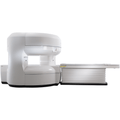
Cone Beam Computed Tomography (CBCT) represents advanced extraoral technology enabling three-dimensional imaging of facial skeleton and dental anatomy. While CBCT systems exceed pure portability requirements due to equipment size, some CBCT manufacturers have developed relatively compact systems suitable for dedicated dental practice installation. CBCT imaging revolutionizes complex surgical planning for dental implants, wisdom tooth extraction, and other surgical procedures requiring three-dimensional anatomic visualization.
Digital Imaging and Diagnostic Advantages
Digital dental radiography provides substantial advantages over traditional film radiography including reduced radiation dose (30-60% reduction compared to film), immediate image availability eliminating film development delays, digital image enhancement enabling diagnostic visualization optimization, and seamless electronic health record integration. These advantages combine to improve practice efficiency, enhance diagnostic capability, and reduce patient radiation exposure—multiple quality improvement benefits from single technology implementation.
Radiation dose reduction in dental radiography is particularly important given that dental patients receive periodic radiographic follow-up throughout their lives (6-month or annual intervals for preventive care, plus additional imaging for treatment planning). Cumulative lifetime dental radiography exposure represents meaningful population-level radiation dose. Digital technology reducing individual radiograph dose by 50% translates to substantial cumulative dose reduction across dentate populations receiving periodic preventive dental care.
Image enhancement capabilities of digital systems enable detection of subtle pathology (early caries, incipient periapical disease) that might not be apparent on film radiographs or grayscale displays. Contrast adjustment, magnification, false-color processing, and other digital enhancements support diagnostic confidence and reduce false-negative findings. These diagnostic advantages improve treatment outcomes and support superior preventive care initiation.
Pediatric Dental Radiography: Special Considerations
Pediatric dental patients present special considerations for radiography due to developing dentition, smaller anatomy, and behavioral/developmental factors affecting patient cooperation. Portable intraoral systems with pediatric-specific positioning aids enable appropriate imaging of developing teeth while minimizing radiation exposure. Panoramic imaging provides complete arch visualization supporting assessment of developing permanent dentition and detection of congenital abnormalities.
Radiation dose minimization in pediatric dental radiography reflects ALARA principles and professional guidelines from American Academy of Pediatric Dentistry (AAPD) emphasizing judicious imaging use justified by clinical indication. Digital systems support dose minimization by enabling lower exposures while maintaining diagnostic quality, particularly important for pediatric patients who may require periodic imaging monitoring throughout development.
Implant Planning and Surgical Guidance
Dental implant therapy has become mainstream treatment option for tooth replacement, requiring comprehensive radiographic assessment for implant site evaluation and surgical planning. Intraoral radiography enables assessment of remaining alveolar bone height at proposed implant sites. Panoramic radiography screens for anatomic landmarks (inferior alveolar nerve, maxillary sinus) that influence implant positioning and require surgical consideration. CBCT imaging enables precise three-dimensional assessment of bone volume, bone quality, and anatomic relationships guiding surgical implant placement planning.
Portable radiography systems supporting implant assessment—intraoral sensors, portable panoramic systems, and potentially CBCT capability—enable comprehensive implant evaluation directly in dental practices. Many dental implant surgeons prefer in-office imaging capability enabling immediate consultation with restorative dentists and streamlined surgical planning coordination. Portable radiography systems supporting implant planning improve practice efficiency and enable dentists to offer comprehensive implant treatment without referring patients to external imaging facilities.
Endodontic Treatment Guidance
Root canal (endodontic) treatment requires radiographic guidance for multiple procedural steps: working length determination (identifying precise root canal length), treatment completion assessment (verifying complete root canal obturation/filling), and post-treatment monitoring for healing and complication detection. Portable intraoral radiography enables rapid radiographic guidance during endodontic procedures, supporting treatment precision and success rates.
Modern digital radiography systems enable real-time image acquisition supporting working length determination with minimal radiation exposure. Electronic apex locators combined with radiographic confirmation enable accurate working length determination while minimizing unnecessary radiographs. Post-treatment radiographs confirm adequate root canal treatment and enable baseline comparison for long-term healing assessment.
Orthodontic Imaging and Treatment Monitoring
Orthodontic treatment planning requires cephalometric radiography and panoramic imaging to assess existing dental and skeletal anatomy, plan tooth movement, and monitor treatment progress. Portable cephalometric systems enable convenient in-office imaging supporting orthodontic treatment planning without requiring patient referral to radiology centers. Serial cephalometric radiographs throughout orthodontic treatment monitor skeletal and dental changes, enabling treatment plan modifications if progress differs from planned trajectory.
Digital cephalometric systems enable computer-assisted treatment planning with automated landmark identification and cephalometric analysis reducing manual measurement burden. Longitudinal digital radiographic records enable automatic comparison of serial cephalometric changes throughout treatment course, objectively documenting treatment progress and outcomes.
Periodontal Disease Assessment
Periodontal disease diagnosis and monitoring requires assessment of alveolar bone levels visualized on radiographs. Bitewing radiographs enable visualization of interproximal bone height supporting periodontal disease classification (gingivitis without bone loss, mild-moderate-severe periodontitis with corresponding bone loss). Panoramic radiographs enable screening assessment of bone loss across entire dentition. Serial radiographs monitor treatment response and disease progression during periodontal management.
Digital radiography enables precise bone level measurement supporting objective periodontal assessment and treatment outcome documentation. Some advanced digital systems incorporate software enabling automated alveolar bone level measurement, reducing manual measurement variability and time burden.
Regulatory Compliance and Radiation Safety
State and federal regulations governing dental radiography require proper credentialing of radiography operators, radiation safety training, equipment quality assurance, occupational dosimetry monitoring, and patient dose documentation. Portable dental systems must maintain equivalent safety standards to fixed installations, with regular quality assurance testing, preventive maintenance, and operator training documentation supporting regulatory compliance.
ARRAD provides dental-specific radiation safety support, quality assurance protocols, and regulatory compliance assistance helping dental practices maintain standards required by state boards, federal agencies, and accrediting organizations like Joint Commission.
Cost-Effectiveness and Return on Investment
Portable dental radiography systems represent capital investment with financial return through improved practice efficiency, enhanced diagnostic capability, and patient satisfaction improvements. Digital technology eliminates film and chemical costs, reducing ongoing supply expenses compared to film-based systems. Rapid image availability reduces appointment time requirements for radiographic procedures, improving practice throughput and revenue generation per operating hour.
Diagnostic quality improvements and enhanced patient communication capabilities from digital systems support improved treatment acceptance rates and reduced patient anxiety, contributing to practice profitability through improved case acceptance and patient loyalty. Many dental practices achieve return on investment for digital portable radiography systems within 3-5 years based on operational efficiency improvements and enhanced revenue generation from improved diagnostic capability.
Integration with Practice Management Systems
Modern portable dental radiography systems integrate seamlessly with practice management software and electronic health records, automating image storage, retrieval, and documentation workflows. Images stored directly in patient records eliminate separate filing systems and improve clinical decision-making efficiency through immediate image availability. Integrated systems support comprehensive patient communication with images displayed during treatment consultation discussions.
Cloud-based digital radiography systems enable access to patient imaging from multiple locations, supporting multi-location practices and enabling remote consultation with specialists or treatment coordinators. Telehealth applications increasingly integrate digital radiography images enabling remote treatment consultation and specialist consultation without requiring patient or clinician travel.
Selecting Dental Radiography Systems for Your Practice
Dental practices evaluating portable X-ray systems should assess practice size, patient demographics (pediatric emphasis, implant focus, etc.), treatment scope (general dentistry vs. specialty), and operational workflow requirements. High-volume practices may prioritize rapid image acquisition and high-throughput capability. Implant-focused practices may prioritize comprehensive imaging including panoramic and advanced capability. Orthodontic practices specifically require cephalometric radiography. ARRAD provides dental-specific consultation helping practices select radiography systems optimized for individual practice characteristics and clinical requirements.
Supporting Dental Excellence Through Diagnostic Imaging
Portable dental radiography systems represent essential infrastructure supporting contemporary dental practice and evidence-based treatment planning. Dental practices committed to diagnostic quality and excellent patient outcomes should prioritize portable radiography system implementation. ARRAD provides dental-specific equipment and support services addressing unique requirements of dental practice.
For dental practitioners seeking to enhance diagnostic imaging capabilities and optimize treatment planning, portable X-ray systems offer proven solutions. Contact ARRAD at 877.299.8303 to discuss portable dental radiography options. Our Lake Forest, California headquarters provides nationwide support for dental imaging systems.